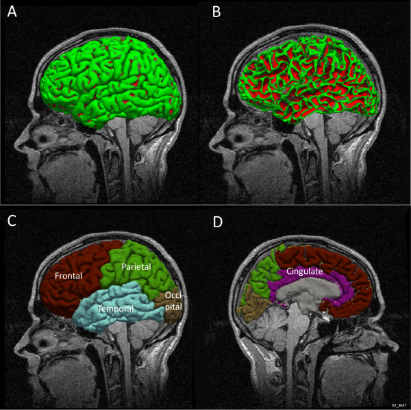
Figure 1.

Lateral visualisation of the pial surface in left hemisphere in one of the subjects (A) and white matter surface in the same subject and position (B). Visualization of the cortical parcellation: external lateral view (C) and mid-sagittal section (D) extracted from FreeSurfer.
