
Figure 1.
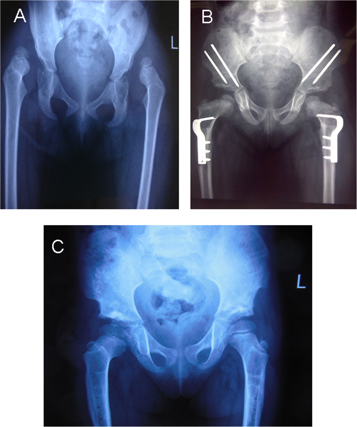
Shows a typical X-ray in a bilateral DDH, both before operation and at the follow up examination. A. Female patient aged 3.8 years with bilateral DDH. Plain X-ray anteroposterior (AP) view. B. Plain X-ray AP view 1 year after one-stage operation, showing good containment of the femoral head. C. AP view 3.9 years postoperatively with excellent clinical and radiographic results.