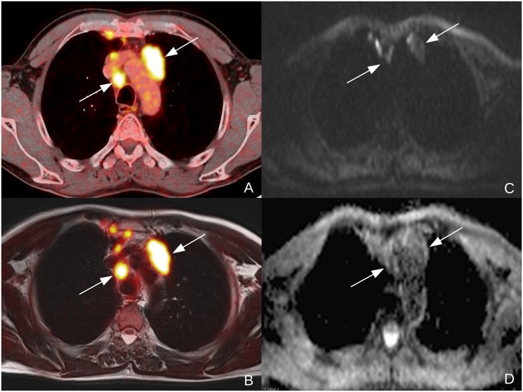
Figure 4. 57y old male patient suffering from NSCLC (tumor stage IIIb, squamous cell carcinoma, unknown tumor grading) Clearly malignant lymph nodes to the left of the aortic arch and right to the trachea in a contrasted enhanced, fused PET/CT-image (A) and in a T2-weighted, fused PET/MR-image (B).
The lymph nodes are clearly depicted in the diffusion weighted b1000-image (C) and show low ADC-values in the corresponding, monoexponential ADC-map. The additional, smaller lymph nodes in the anterior mediastinum were not visible in DWI (C) and were therefore excluded from further analysis.

