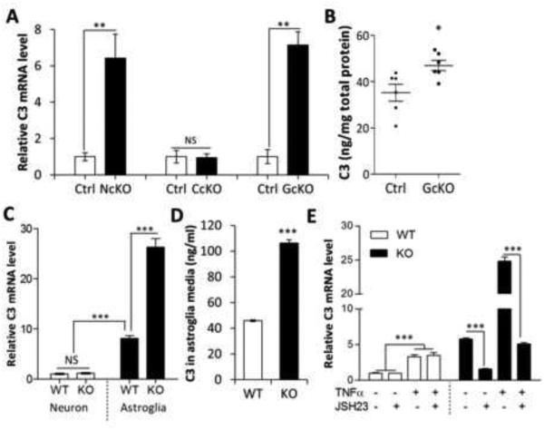
Figure 1. C3 is overexpressed in IκBα-deficient astroglia.
(A) Quantitative RT-PCR measurement of C3 mRNA expression in hippocampal samples of 2-month-old Nestin-Cre; IκBαfl/− (NcKO), CamKIIα-Cre; IκBαfl/− (CcKO), and GFAP-Cre; IκBαfl/− (GcKO) mice and their littermate controls (Ctrl). (B) C3 protein levels in GcKO and Ctrl hippocampi measured by ELISA. (C) C3 mRNA levels in wild-type (WT) and IκBα knockout (KO) primary neurons or astroglia. (D) ELISA quantification of C3 protein levels in conditioned media of WT or IκBα KO astroglial cultures. (E) C3 mRNA expression in WT or IκBα KO astroglial cultures treated with different combinations of TNFα (50 ng/ml) or NFκB inhibitor JSH23 (20 μM). N=3 per group per experiment except N=6 in (B). A, B, and D: Student’s t-test; C: Two-way ANOVA followed by pairwise comparison. E: Three-way ANOVA followed by pairwise comparison. *P < 0.05; **P < 0.01; ***P < 0.001; NS: non-significant. See also Figure S1.

