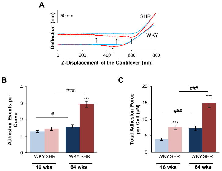
Figure 3.
Adhesion properties of VSMCs are increased in SHR. In separate atomic force microscopy experiments, the microcantilever probe was coated with fibronectin, to promote the formation of VSMC surface attachments to the probe. This resulted in several stepwise adhesion breaks (arrows), as the cantilever was pulled away from the cell surface, as shown for representative approach (blue) and retraction (red) curves (A). SHR cells formed a greater number of adhesion attachments per approach-retraction cycle (B). The total force generated from these adhesion attachments was also greater in the SHR (C). Data are shown as mean ± SEM, with n = 60 cells per group. Post-hoc comparisons, * p < 0.05, ** p < 0.01, **** p < 0.0001 compared to WKY; # p < 0.05, ## p < 0.01, ### p < 0.001 compared to young.

