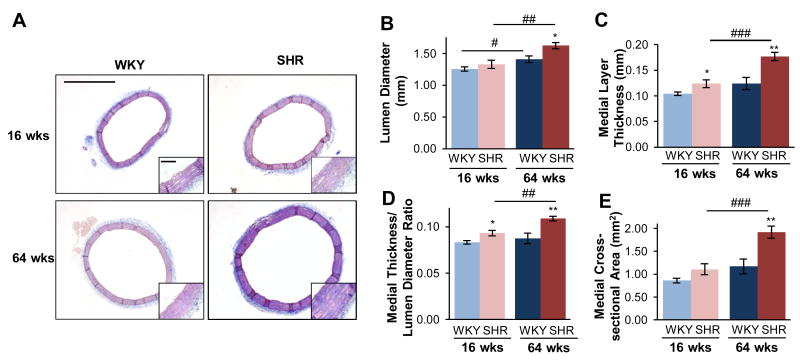
Figure 4.
Aortic structural remodeling in hypertension differs from that of age. Overall morphology was compared from Mason's Trichrome staining of the thoracic aorta (scale bar = 1.0 mm), shown with high-magnification insert (scale bar = 0.1 mm) (A). At both ages, the SHR had increased lumen diameter (B), medial layer thickness (C), and medial thickness-diameter ratio (D), and medial cross-sectional area (E), and these changes further increased with age. Data are shown as mean ± SEM, with n = 8 animals per group. Post-hoc comparisons, * p < 0.05, ** p < 0.01, compared to WKY; # p < 0.05, ## p < 0.01, ### p < 0.001, compared to young.

