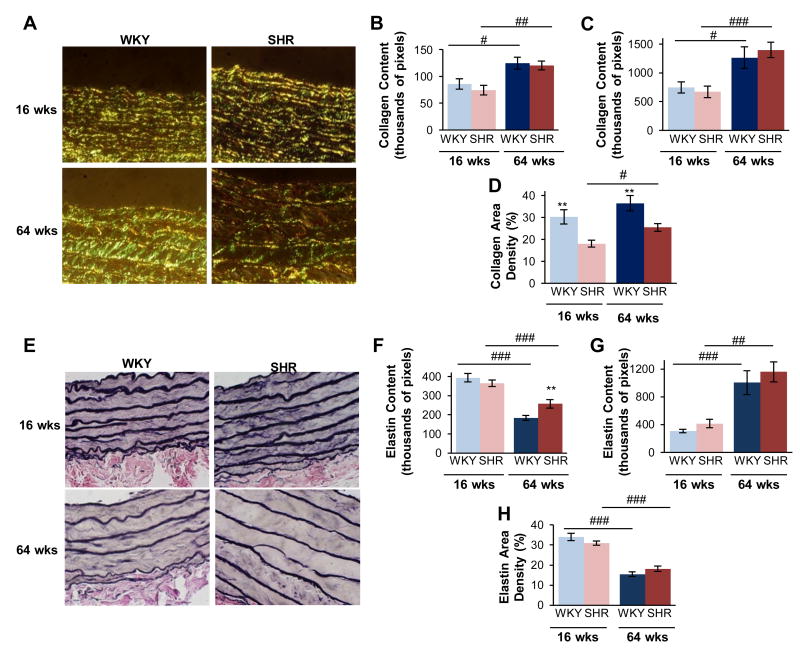
Figure 5.
Collagen and elastin composition in hypertension differs from that of age. Collagen fibers were visualized by under circularly-polarized light (A), and neither the content within individual image fields (B) nor that computed for the total tissue (C) was not significantly different in hypertension. The collagen content was normalized to the tissue area within that field to determine a tissue density, which was decreased in the SHR (D). Elastin was quantified from Van Gieson's staining (E). Older age decreased elastin content within individual fields (F), but overall this was increased for the total tissue ring(G). Elastin density was decreased by age, and not hypertension (H). Data are shown as mean ± SEM, with n = 8 animals per group. Post-hoc comparisons, * p < 0.05, ** p < 0.01, compared to WKY; # p < 0.05, ## p < 0.01, ### p < 0.001, compared to young.

