Figure 5.

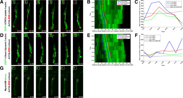
The primary cilium is embedded in the actomyosin network during the two-stroke motility cycle. The primary cilium (labeled by Arl13b-KO1, red) is embedded within (A) the stable F-actin (labeled by EGFP-UTRCH, green), (D) the dynamic F-actin (labeled by EGFP-Lifeact, green), and (G) the myosin ii heavy chain-labeled (labeled by MHCiiB-Venus, green) contractile domains in the leading process of living neurons. (B, E) Adaptive volumetric kymographs of the sequences shown in Panel A and D (somal boundaries, dashed blue line; soma center, solid blue line; cilium position, red line; EGFP-UTRCH or EGFP-Lifeact, green). (C, F) Analysis of mean actin cilium computed within a 1, 2, or 4 μm radius from the center of the cilium (black dashed line represents the average actin concentration throughout the cell). Scale bar, 10 μm. Inset shows the extent of co-localization at each time point. Scale bar, 10 μm.
