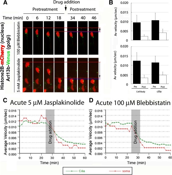
Figure 6.

Myosin ii and F-actin dynamics motor activity are required for coordinated movement of the cilia and cell body. CGNs were induced to express the cilia label Arl13b-Venus (green) and the nucleus label H2B-mCherry (red), and time-lapse imaging was used to measure velocity of the cilia and soma in migrating neurons. After migration for 18 minutes, 100 μM blebbistatin or 5 μM jasplakinolide were added, and imaging continued for a further 28 minutes. Representative time-lapse images are shown. (A) Both drugs potently inhibited forward movement. (B) Velocity of the cell body and cilia before and after drug treatment; 100 μM blebbistatin (upper) reduced mean cell body velocity from 0.009 ± 0.004 to 0.002 ± 0.001 μm/sec (n = 191) and reduced mean cilia velocity from 0.011 ± 0.004 to 0.004 ± 0.002 μm/sec (n = 163); 5 μM jasplakinolide (lower) reduced mean cell body velocity from 0.012 ± 0.004 to 0.003 ± 0.002 μm/sec (n = 128) and reduced mean cilia velocity from 0.011 ± 0.004 to 0.005 ± 0.002 μm/sec (n = 143). All changes in velocity were statistically significant (P <0.0001, t-test). (C, D) Decrease in mean velocity at each time point after addition of (C) 5 μM jasplakinolide (n = 143 cilia, 128 somas) (see movie) and (D) 100 μM blebbistatin n = 163 cilia, 191 somas).
