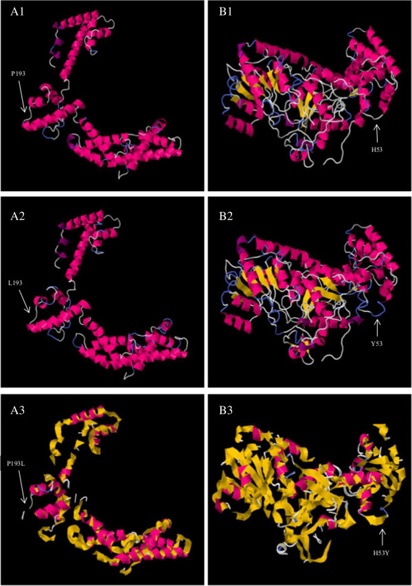
Figure 5.

Modelled protein structures of RpoS and TviE. A1 showed native RpoS with proline at position 193. A2 showed mutant RpoS with amino acid leucine at position 193. A3 showed superimposed structure of RpoS native structure (yellow) with mutant structure (pink). B1 showed native TviE with histidine at position 53. B2 showed mutant TviE with amino acid tyrosine at position 53. B3 showed superimposed structure of TviE native structure (yellow) with mutant structure (pink).
