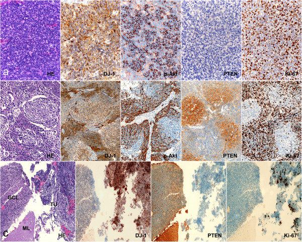
Figure 2.

Immunohistochemical analysis of DJ-1, PTEN, p-Akt, and Ki-67 expressionin MBs. (a) In classic MBs, DJ-1 and p-Akt appeared to have diffused positive signal in tumor cells, but PTEN was negative in most cases. Proliferative activity (Ki-67 labeling) was particularly prominent in areas with high expression of DJ-1 and p-Akt. (b) In desmoplastic MBs, high expression of DJ-1, p-Akt, and Ki-67 was observed in internodular areas with undifferentiated or poorly differentiated tumor cells; however, detectable signals of PTEN were observed in intranodular areas with neuronal differentiation. (c) Immunohistochemical analysis showed that expression of DJ-1 was significantly higher in tumor cells than in normal cerebellum, but PTEN was found to be expressed at significantly lower levels in tumor cells compared with adjacent normal cerebellum. ML, molecular layer of cerebellum; GCL, granule cell layer of cerebellum; TU, tumor. (Original magnification × 200).
