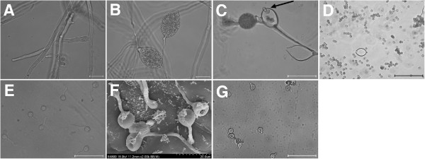
Figure 1.

Life cycle stages of P. cactorum including cysts germinating under different conditions. Five successive life cycle stages are shown: (A) mycelia, (B) sporangia, (C) released sporangia and zoospores, (D) cysts, (E)-(G) germinating cysts. In panel (C), the arrow indicates a zoospore being released from the sporangium. Cysts were germinated on a cellophane membrane placed on the top of an N. benthamiana leaf (E), or directly on N. benthamiana leaves (F), or in water (G). In panel (F), the cysts were observed under a Cryoscanning electron microscope (Hitachi S-4800 SEM). The other cell types were observed using an Olympus System Microscope BX53. Photos were taken at 70 (E), 60 (F) and 300 (G) min post-inoculation. Scale bars: (A), (B), 10 μm; (C), (E), (G), 50 μm; (D), 100 μm.
