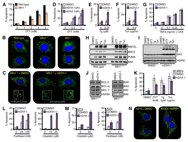
Figure 3. Mitochondrial network shape regulates tUPR.
(A) Wt and Mfn1−/− MEFs were treated with DTT for 18 h. (B) Wt, Mfn2−/−, and Mfn1−/− MEFs were loaded with MitoTracker Green® (50 nM) and Hoechst 33342 (20 μM) before imaging (400×). (C) Mfn1−/− MEFs were treated with mDIVI-1 (25 μM) for 2 h before imaging (400×). Further magnified regions (2.5×) are shown in white boxes. The average length of ~ 200 mitochondria is shown. (D) Mfn1−/− MEFs were pre-treated with mDIVI-1 (25 μM) for 2 or 8 h, and then DTT for 18 h. (E-F) Mfn1−/− MEFs were pre-treated with mDIVI-1 (25 μM) for 8 h, then Tg (0.25 μM) or Tun (0.5 μg/ml) for 18 h. (G) Mfn1−/− MEFs were pre-treated with mDIVI-1 (25 μM) for 8 h, then TNFα and CHX (10 μg/ml) for 18 h. (H) HM fractions from ER stress treated Mfn1−/− MEFs were analyzed by western blot. (I) Mfn1−/− MEFs were pre-treated with mDIVI-1 (25 μM) for 2 h, ER stress agents for 18 h, and mitochondria were isolated and analyzed by western blot. High molecular weight complexes of BAX are indicated (*). VDAC is a loading control. (J) Mfn1−/− MEFs were treated with mDIVI-1 (25 μM) for 8 h, and lysates were analyzed by western blot. (K) Wt MEFs were pre-treated with mDIVI-1 (25 μM) for 8 h, and ER stress agents for 18 h. (L) Mfn1−/− MEFs were pre-treated with mDIVI-1 (25 μM) for 8 h, and Paclitaxel or Cisplatin for 18 h. (M) Same as L, but A375. (N) Same as C, but A375. All data are representative of at least triplicate experiments, and reported as ± S.D., as required. See also Figure S4.

