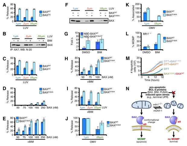
Figure 7. BAX α9 displays requirements for membrane shape.
(A) LUVs were combined with BAX or BAXOG (0.25 μM) for 15 min at 37°C. (B) BAX (100 ng) was incubated in the presence of BIM BH3 (2.5 μM) and LUVs for 30 min prior to 6A7 IP and western blot. (C) LUVs were combined with BAXWT or BAXΔC (0.25 μM) for 1 h at 37°C. The required incubation time is longer for BAXΔC compared to BAXWT, which increases BAXWT activity. (D) LUVs were combined with BAXWT or BAXS184A for 30 min at 37°C. (E) Same as D, but with BIM BH3 (2.5 μM). (F) LUVs were combined with BAXWT or BAXS184A (100 nM) for 30 min at 37°C prior to centrifugation, solubilization, and western blot for associated BAX. (G) NBD-BAXWT or NBD-BAXS184A was incubated with 1 μm LUVs for 5 min, ± BIM BH3 (2.5 μM). An increase in NBD fluorescence indicates BAX·LUV interactions, and is reported as fold increase compared to NBD-BAXWT + LUVs. (H) LUVs (1 μm) were combined with BAXWT or BAXS184A (50, 75, 100 nM) with BIM BH3 (2.5 μM) for 30 min at 37°C. (I) LUVs were combined with BAXWT or BAXS184A (50 nM) with BIM BH3 (2.5 μM) for 30 min at 37°C. (J) OMVs were combined with BAXWT or BAXS184A (50 nM) and BIM BH3 (2.5 μM) for 30 min at 37°C. (K) NBD-BAXWT or NBD-BAXS184A ± BIM BH3 (2.5 μM) was incubated with OMVs for 30 min at 37°C. The interaction between NBD-BAXWT + BIM BH3 with 1 μm OMVs is reported as 100%. (L) Digitonin-permeabilized, JC-1 loaded Mfn1−/− MEFs were incubated with BIM BH3 (0.1 μM), BAXWT (50 nM), and BAXS184A (50 nM), and ΔΔψM was determined. (M) Mfn1−/− MEFs expressing shBax were reconstituted with human BAXWT or BAXS184A, treated with DTT (1.5 mM), and the kinetics of tUPR was evaluated by IncuCyte. (N) A schematic summarizing the relationship between BAX, mitochondrial shape, and apoptosis. All data are representative of at least triplicate experiments, and reported as ± S.D., as required. See also Figure S7.

