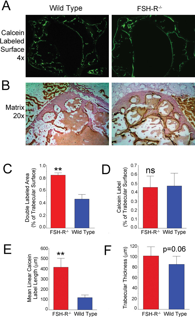
Figure 1. FSH-R–mediated effects on bone formation and bone degradation.
(A). Whole vertebral cross section of the Fshr−/− mouse calcein labeled 5 and 1 d before sacrifice. In these 3 mm fields, the wild-type mouse (left) has typical short regions of labeling, while the knockout (right) shows a unique pattern with long regions of active bone formation. (B). Hematoxylin-stained cross sections at 20× (fields are 600 µm across) show typical variability in the wild-type (left), but uniform and smooth swiss cheese–like trabecular bone in the Fshr−/−. (C). A much greater proportion of bone was double labeled in the FSH-R−/− animal (p < 0.01). (D). Total calcein label was unchanged. (E). Linear cross-sectional length of calcein labeling was increased in the FSH-R−/− animal (p < 0.01), reflecting the size of bone forming units. (F). Trabecular thickness was increased in the Fshr−/− consistent with previous work,2 but had marginal statistical difference (p = 0.06) in this case.

