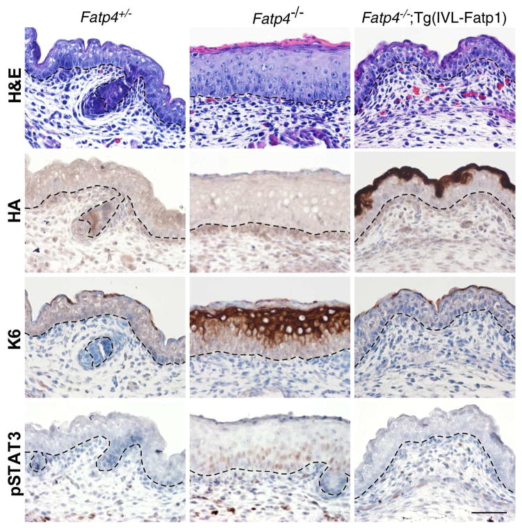
Figure 2. Amelioration of skin phenotypes in Fatp4−/−;Tg(IVL-Fatp1) mice.
Dorsal skin sections from E16.0 embryo littermates were subjected to hematoxylin and eosin staining (top row) and immunohistochemical analyses followed by counterstaining with hematoxylin (bottom three rows). The thickened epidermis phenotype seen in Fatp4−/− newborns was normalized in Fatp4−/−;Tg(IVL-Fatp1) newborns (top row). The HA-tagged FATP1 encoded by the transgene was detected primarily in granular keratinocytes in Fatp4−/−;Tg(IVL-Fatp1) mice (second row). The ectopic expression of keratin 6 seen in Fatp4−/− newborns was diminished in Fatp4−/−;Tg(IVL-Fatp1) mice (third row). The nuclear localization of pSTAT3 shown in Fatp4−/− newborns was also diminished in Fatp4−/−;Tg(IVL-Fatp1) mice (bottom row). Dashed lines demarcate the dermo-epidermal boundary. Scale bar is 50 μm.

