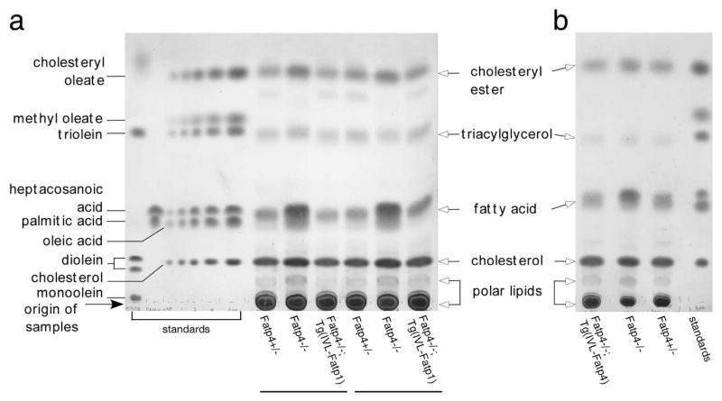
Figure 3. Amelioration of the elevated level of free fatty acids in Fatp4−/−;Tg(IVL-Fatp1) and Fatp4−/−;Tg(IVL-Fatp4) epidermis.
Free, extractable lipids from the epidermis of newborn mice were resolved by TLC and visualized by charring the plate. The elevated level of free fatty acids seen in Fatp4−/− mice was reduced to normal in Fatp4−/−;Tg(IVL-Fatp1) (a) and Fatp4−/−;Tg(IVL-Fatp4) mice (b). Components of the lipid standards and origin of sample application are indicated on the left of a. Lipid species of the epidermis are indicated between the panels.

