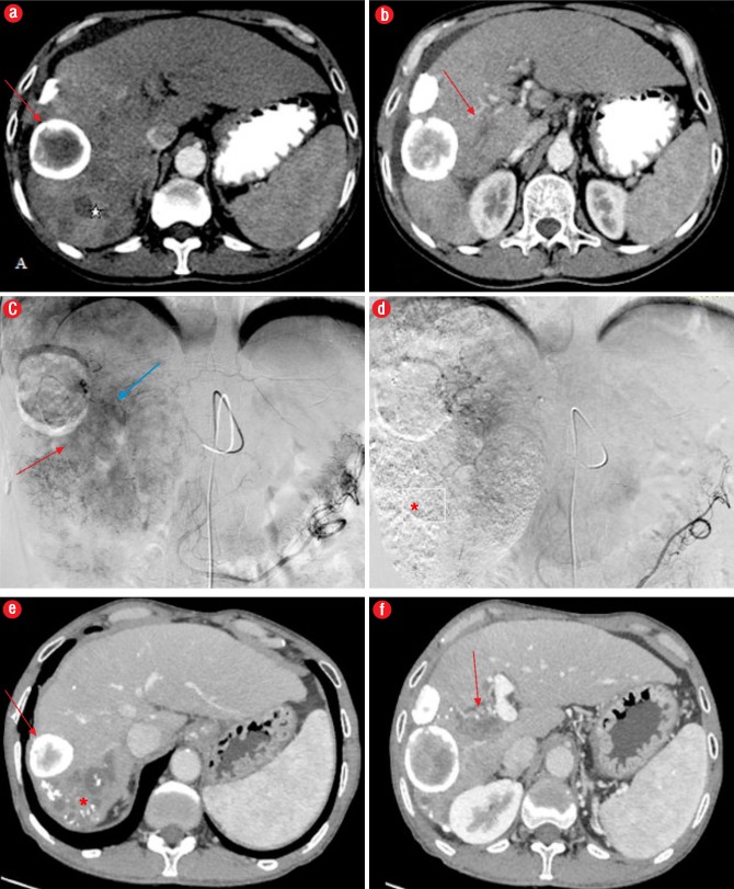
Figure 3.
Computed tomography (CT) angiography images from a 62-year-old male patient with right portal vein (PV) thrombosis; (a) CT indicates areas of arterial enhancement* representing HCC, and a peripherally calcified lesion anterior to HCC suggestive of a calcified hydatid (red arrow); (b) Right PV thrombosis (red arrow); (c) Catheter angiography image showing areas of tumoral blush (red arrow) and enhancing tumor thrombus in PV (blue arrow); (d) Post- embolization image showing no residual blush in the tumor with lipidiol deposition (*); (e, f) CT angiography after one year shows absence of arterial enhancement with lipidiol deposition(*) and non-enhancing PV thrombus (red arrow).

