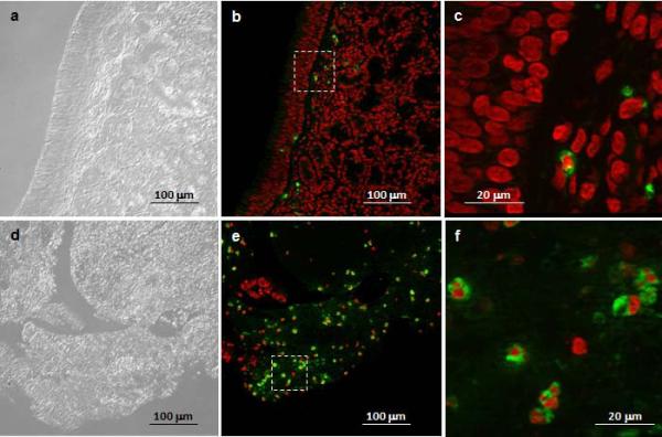
Figure 1. Quantification of neutrophils in sinus tissue from non-CRS and CRS subjects.
A. Neutrophils were detected with polyclonal antibodies against HNP1-3 and visualized with Alexa Fluor 488 labeled secondary antibodies, and tissues were counterstained with the nuclear stain propidium iodide. Alexa-Fluor 488 stain demonstrates HNP-positive neutrophils in green color, and propidium iodide highlights nuclei in red color. Shown are representative images for non-CRS (top, a-c) and CRS without nasal polyposis (bottom, d-f). a, d: phase contrast; b, c, e, f: overlay for Alexa-Fluor 488 and propidium iodide. The squares in b and e represent the area shown in c and f, respectively. B. Enumeration of neutrophils in sinus tissue sections from non-CRS and CRS subjects. Shown are means ± SEM, n = 4 for non-CRS and n = 13 for CRS. All CRS cases were without nasal polyposis. *p = 0.010 in independent samples Mann Whitney U test. HNP: human neutrophil peptide; CRS: chronic rhinosinusitis

