
Figure 2.
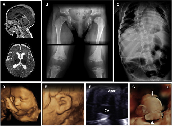
Radiographic Features of CODAS Syndrome
(A) Sagittal T1 (upper) and axial T2 (lower) images of an affected 4-year-old Amish child show mild diffuse cortical atrophy, an immature pattern of myelination, and hypoplasia of the corpus callosum.
(B) Skeletal radiographs show metaphyseal dysplasia most evident at hip joints (upper) and valgus knees with both hypoplasia and delayed ossification of epiphyses (lower).
(C) Severe scoliosis is evident early in childhood, and coronal clefts are observed at various levels of the vertebral column.
(D–G) Prenatal imaging of a 32-week-old fetus with CODAS syndrome revealed polyhydramnios, a two-vessel umbilical cord, midfacial hypoplasia, (D) crumpled helices, (E) a balanced atrioventricular canal, (F) common atrium, omphalocele, (G, arrow) and absence of left lower long bones, which were replaced by a relatively well-formed foot attached directly to the hip. (G, arrowhead) The asterisk in (G) marks the left palpebral fissure for orientation.