
Figure 7.
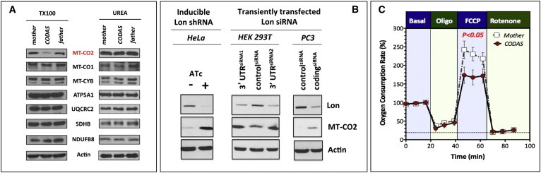
Alterations in the Structure and Function of the Electron Transport Chain in Proband LCLs
(A) For extraction of cellular proteins, lysis buffer containing 0.1% Triton X-100 (TX100) or 8 M urea (UREA) was used. Immunoblotting was performed for proteins as shown. MT-CO2 abundances were selectively reduced in CODAS cells but were recovered in the presence of 8 M urea.
(B) MT-CO2 is a protein substrate of mitochondrial Lon. A HeLa cell line with a stably integrated anhydrotetracycline (ATc)-inducible shRNA targeting Lon was grown in the presence or absence of ATc (500 ng/ml) for 4 days. HEK293T cells and PC3 cells were transiently transfected with a siRNA targeting the 3′ untranslated regionof Lon or a control siRNA and cultured for 3 days. Cell extracts (25 μg) were immunoblotted for Lon, MT-CO2, or actin as the loading control.
(C) Basal normalized oxygen-consumption rate was established at 18 min (representing 100%), followed by addition of oligomycin (Oligo, 1 μM); FCCP (750 nM); and finally rotenone (1 μM). A Student’s t test showed that cells from CODAS-syndrome-affected probands had significantly (p < 0.05) lower spare mitochondrial respiratory capacity in the presence of the uncoupler FCCP. Error bars represent ±SD.