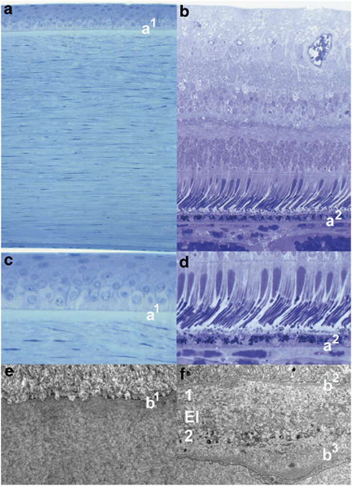
Figure 3.
Light microscopy of the human cornea and retina. (a and b) The locations of Bowman's (a1) and Bruch's (a2) membranes seen in a higher power in (c and d), respectively. Transmission electron micrographs of Bowman's (e) and Bruch's (f) showing the basement membrane of the corneal epithelium (b1) overlying a fine matrix of collagen fibrils in (e), while the basement membranes of retinal pigment epithelium (b2) and the endothelial cells of the choriocapillaris (b3) are seen in (f). The inner and outer layers of Bruch's membrane are labelled 1 and 2, respectively, and are separated by the elastin layer El in (f).

