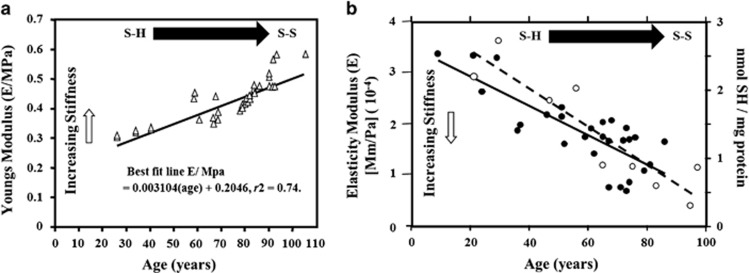
Figure 8.
Graphs showing the change in elasticity as a function of age in the cornea (a) and Bruch's membrane (b). In the cornea, the change is measured in terms of Young's modulus of elasticity as it is thought to reflect changes in the bond structure of the tissue with age, particularly the collagen. The reversal of the slope seen in (b) is as a consequence of measuring elastic moduli and not Young's modulus (open circles). In the retina, it was possible to use a fluorescent technique to determine quantitatively the decrease in the number of fluorescent SH bonds, which are flexible and gave a measure of the non-fluorescent rigid SS bonds (closed circles).

