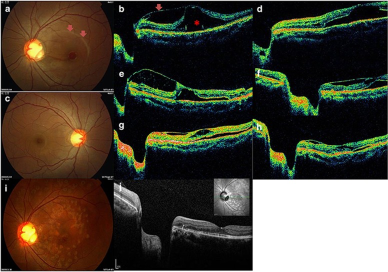
Figure 1.
Composite of representative clinical findings from patient 1. Preoperative fundus findings in the left eye. A 1/5 PD yellowish depression at the infratemporal optic disc with serous retinal detachment involving the macula (arrow) is visible (a). C/D ratio in both eyes (a, c) is about 0.7. Visual acuity in the affected left eye at presentation was 20/200. Preoperative horizontal OCT in the left eye (b) demonstrated partial vitreomacular traction (arrow) and serous detachment (*) involving the macula. The intra-retinal fluid was most prominent at the level of the outer plexiform layer with some vertical bridging tissue spanning a schisis-like cavity. OCT 10 days postoperatively (d) showed marked reduction of the schisis-like cavity. However, the cavity and serous retinal detachment still existed 1 month postoperatively (e). C3F8 intravitreal injection and photocoagulation was repeated. A significant anatomical improvement was achieved, and the visual acuity gradually improved to 20/40 2 weeks after the 2nd injection (f). Marked absorption of intra-retinal and sub-retinal fluids was noted by OCT 2 months after the repeat injection (g). OCT demonstrated a retinal thickness of 268 μm 4 months' after repeated treatment (h) versus 744 μm (b) before treatment. No recurrence or visual field defects were noted during the follow-up period. Fundus photograph and OCT are shown in i 8 months after repeated injection. The last OCT (Spectralis) at 50 months' follow-up is shown in j.

