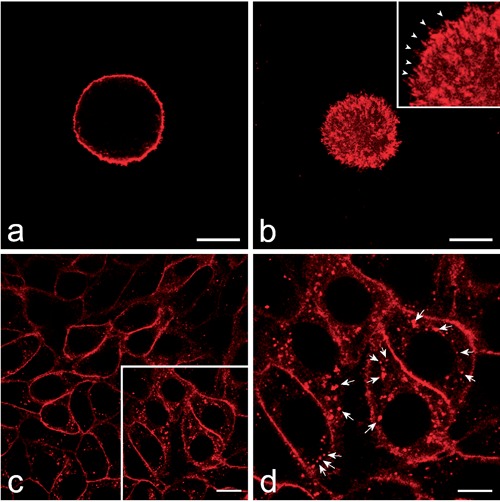
Figure 1.

Confocal optical sections of HeLa cells; the labelling with red fluorescing PKH26 was performed on cells either in suspension (a,b) or adhering to the glass cover-slip (c,d). a) Equatorial optical section of a HeLa cell showing the plasma membrane labelling. b) Superficial optical section of the same cell whose tiny membrane protrusions are also finely labelled (arrowheads in the inset). The plasma membrane fuorescence is apparent as well after labelling of adhering cells (c) in which brightly fluorescing spots are also present inside the cytoplasm. d) The higher magnification of the frame in c allows a better visualization of the intracellular fluorescing spots (arrows). Scale bars: 20 µm.
