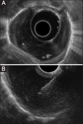
Figure 1.

Endoscopic ultrasound findings. (A) A homogenous hypoechoic well-circumscribed tumor with some calcifications inside, originating from muscle layer and that completely encircles the upper third of the esophagus. (B) Endoscopic ultrasound-guided fine-needle aspiration of the tumor
