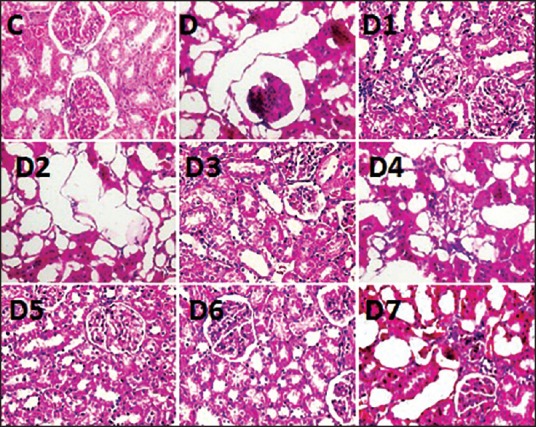
Figure 1.

H&E staining of kidney structure containing tubules, interstitium and glomerulus. H&E staining of kidney structure containing tubules, interstitium and glomerulus. C - control with normal tubules, interstitium and glomerulus. D - diabetic control with extensive tubular dilatation showing epithelial cell atrophy severe interstitial inflammations (not shown) and normal glomerulus. D1, D3, D6 shows moderate tubular dilatation with mild to moderate interstitial inflammations (not shown) and normal glomerulus. D5 photograph shows only mild tubular dilatation with mild interstitial inflammations and normal glomerulus. (magnification 400×)
