Figure 2.

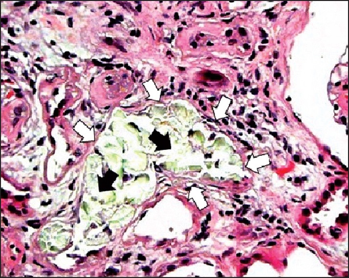
Histopathology (hematoxylin-eosin; original magnification, ×400); Renal tubular dilation (white arrows) with calcium oxalate (CaOx) deposits (black arrows) in group B

Histopathology (hematoxylin-eosin; original magnification, ×400); Renal tubular dilation (white arrows) with calcium oxalate (CaOx) deposits (black arrows) in group B