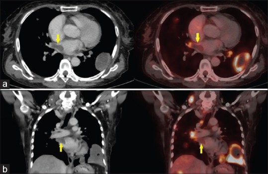
Figure 3.

18F-fluoro-deoxyglucose (FDG) contrast enhanced positron emission tomography-computed tomography (PET-CT) images. (a) Transaxial CT and corresponding fused PET-CT, (b) coronal CT and corresponding fused PET-CT sectional images showing non-FDG avid thrombus in right superior pulmonary vein extending upto the left atrium (arrow)
