
Figure 1.
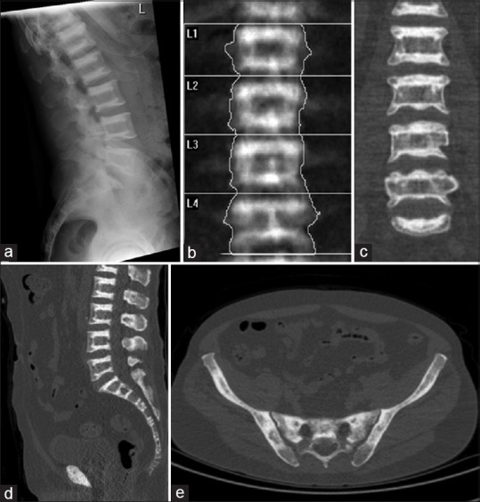
(a) X-ray of spine shows dense and sclerosis at the margins of the vertebral bodies in alternating parallel sclerotic and lucent bands (sandwich vertebrae or “rugger-jersey” spine). (b) Dual-energy X-ray absorptiometry scan shows dense sclerosis at the margins of the vertebral bodies and the T-score was high at +6.5 (b and c). On computed tomography (CT) component of single-photon emission computed tomography/CT, there is dense sclerosis at the margins of the vertebral bodies (bone-in-bone appearance) and within the pelvic bones (d and e)