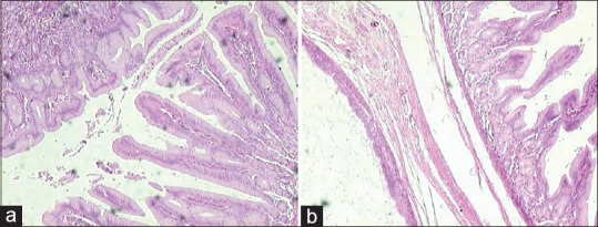
Figure 3.

Cyst bound by a layer of smooth muscle and fibroconnective tissue and lined by pseudostratified ciliated columnar epithelium (respiratory; left side) along with the glandular epithelium comprising of foveolae (pits) and gastric glands (gastric mucosa; right side) (a; H and E stain, ×40). Areas from the cyst showing well developed gastric mucosa (b; H and E stain, ×40)
