Figure 2.

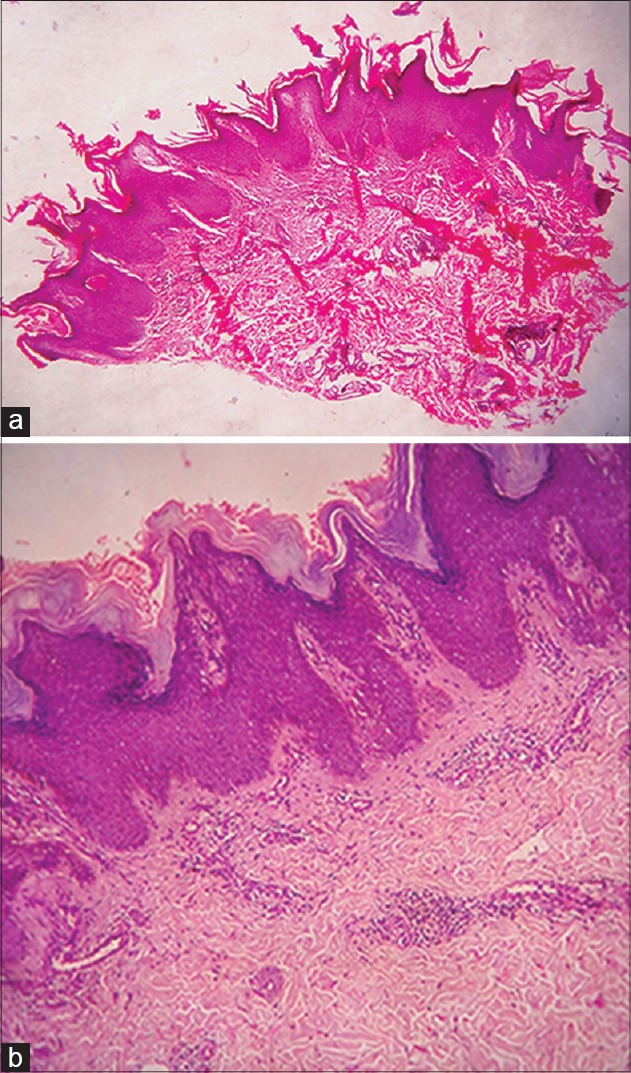
(a) Hyperkeratosis, acanthosis, papillomatosis and sparse mononuclear cell infiltration in dermis (H and E, ×40) (b) Higher magnification (H and E, ×100)

(a) Hyperkeratosis, acanthosis, papillomatosis and sparse mononuclear cell infiltration in dermis (H and E, ×40) (b) Higher magnification (H and E, ×100)