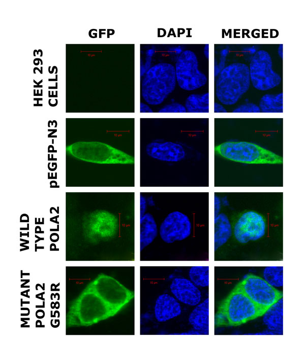
Figure 3.

Subcellular localization of GFP-tagged POLA2 wild type and mutant G583R proteins. HEK 293 cells were transfected with GFP-fused proteins (green) as indicated and treated with anti-GFP followed by Alexa 488 (green) to stain the proteins and 4',6-diamidino-2-phenylindole (DAPI) (blue) to stain the nuclei and then examined by laser fluorescence confocal microscopy. The fields shown were visualized independently at the appropriate wavelength for anti-GFP (488 nm) and DAPI (405 nm), and then the two images were merged. Magnification: 63×. Scale bar is 10 µm.
