
Figure 7.
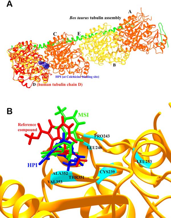
(a) Depiction of the complete Bos taurus tubulin assembly comprising chains A, B, C, D and E of which A and C belong to the α chain (shown in orange); B and D belong to the β chain (shown in yellow). The modelled human tubulin β-chain has been compared (in red) and the ligand is shown in blue bound at the α-β interfacial colchicine binding site. (b) Depiction of the binding modes of HPI (in blue) and MSI (in green) compared to the most active arylthioindole derivative (in red) taken as the reference compound and important residues (in cyan).