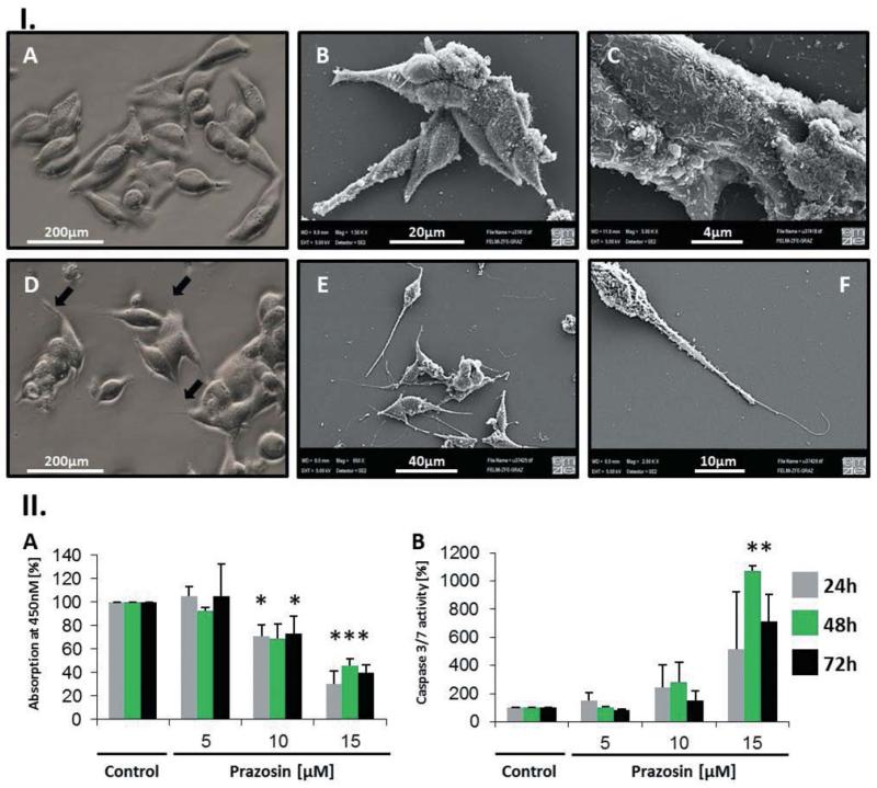
Figure 2.
Prazosin induced morphological alterations and apoptosis in the TT cell line. I. A: Light microscopic view of TT cells: Typical TT cells are small, spindle-shaped and growing in small clusters. B/C: Scanning electron microscopic (SEM) analysis of TT cells exhibited a highly complex cytoplasmic membrane showing many membrane-bound vesicular structures, which may reflect exocytotic processes. D: Light microscopic view of 24 h prazosin (15 μM)-treated TT cells: Prazosin-treated cells were generally more spindle-shaped and exhibited long filopodia like polar fibres (arrows). E/F: SEM analysis of 24 h prazosin-treated cells (15μM) confirmed the enhanced spindle-like character of TT cells upon prazosin treatment and showed that the formed fibres are very fragile structures. The fibres seem to protrude from the polar endings of the cells (F). II: A: Growth, respectively viability, of TT cells was tested using the WST-1 reagent over 72 h. A dose-dependent decrease of the optical density (OD) at 450 nm was evident when prazosin was added to the culture; n=4. B: Activation of effector caspases 3/7 indicates that prazosin induces apoptosis in TT cells; n=3. *p<0.05 according to one-way ANOVA with the Holm-Sidak post hoc test.

