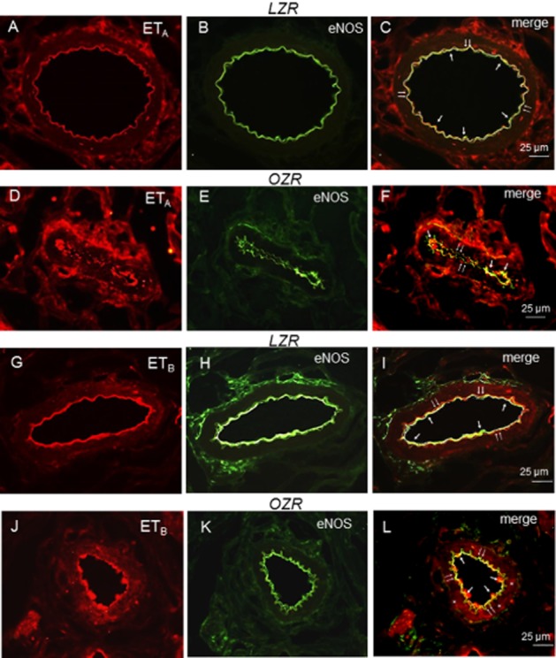
Figure 9.

Representative immunohistochemical staining of ETA and ETB receptor expression in the endothelium and smooth muscle layer of penile arteries from LZR and OZR. (A, D) Immunofluorescence for ETA receptors (red areas) is distributed in the endothelial and media layers of the arterial wall in LZR and OZR. (B, E) Endothelial cell layer was visualized with the anti-eNOS marker (green). (C, F) Immunofluorescence double labelling for eNOS marker and ETA receptor expression in endothelial cell layer demonstrates colocalization in endothelium (yellow areas) in LZR and OZR penile arteries. (G, J) Immunofluorescence for ETB receptors (red areas) is distributed in the endothelial layer of the arterial wall in LZR and OZR penile arteries and in the smooth muscle layer in OZR (asterisks). (H, K) Endothelial cell layer was visualized with the anti-eNOS marker (green). (I, L) Immunofluorescence double labelling for eNOS marker and ETB receptor expression in endothelial cell layer demonstrates colocalization in endothelium (yellow areas, arrows) in LZR and OZR. Scale bars indicate 25 μm. Sections are representative of n = 3 LZR animals and OZR animals. Double arrows: internal elastic layer.
