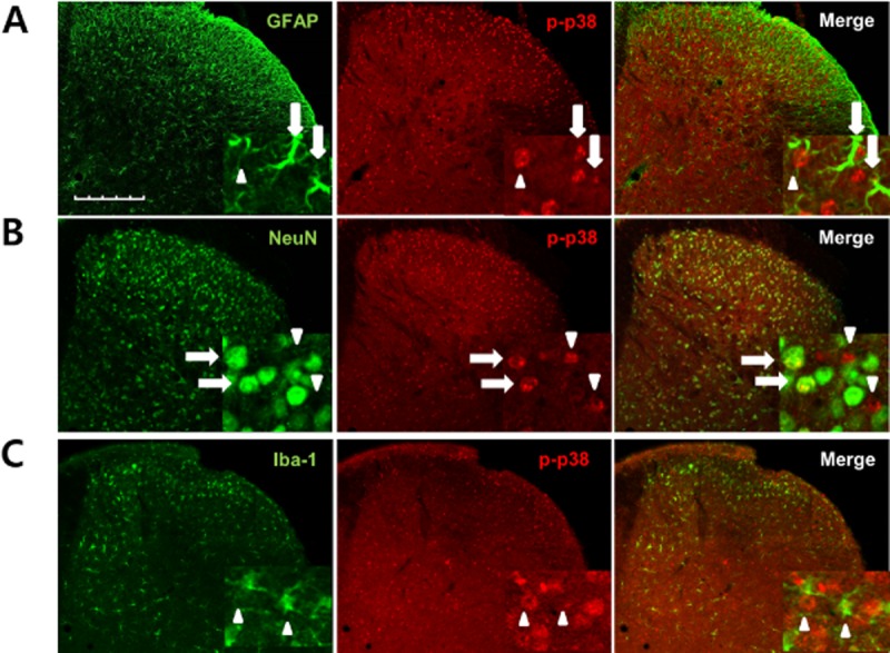
Figure 5.

Phosphorylated (p)-p38 was increased in spinal astrocytes and neurons following CCI in mouse ipsilateral spinal cord dorsal horn. (A–C) Transverse sections through the lumbar spinal cord segment 3 days after CCI were labelled with an antibody against p-p38 (red) and double labelled with GFAP, Iba-1 or NeuN antibodies (green). The p-p38-ir staining was preferentially located in the nuclei of GFAP-positive and NeuN-positive cells. (A) Some of the p-p38-labelled cells were GFAP-positive astrocytes (arrows), while others were not GFAP positive (arrowheads). (B) p-p38-labelled cells were clearly double labelled with NeuN antisera, a neuronal marker (arrows), although some phospho-p38-labelled cells were not NeuN-positive neurons (arrowheads). (C) No colocalization was detected for p-p38 and Iba-1, a microglia marker (arrowheads). Scale bar, 200 μm.
