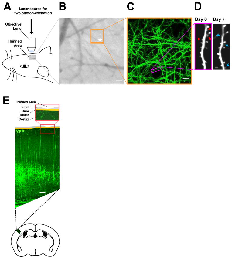
Figure 1. Live imaging studies of spine dynamics of YFP-labeled layer V pyramidal neurons in the primary somatosensory cortex.
(A) Schematic of transcranial in vivo imaging based on two photon-excitation laser scanning microscopy using a pulsed, mode-locked Ti:sapphire laser light source.
(B) CCD camera view of the cortical vasculature beneath a thinned-skull window in a YFP-expressing mouse. The region outlined in orange indicates the area where low- and high-magnification images of dendritic branches were obtained in repeated live imaging. The white arrow points to the tip of a vessel used to relocate the imaged region. Scale bar, 50 μm.
(C) Two-dimensional projection of the low-magnification three-dimensional image stack of dendritic branches and axons from the boxed region in B. A black circular area devoid of YFP (green)-labeled processes in the lower center is the tip of the vessel shown in the boxed region in B. Scale bar, 10 μm.
(D) High-resolution images of a YFP-labeled dendritic segment (box in C) acquired on Day 0 and Day 7. Red arrows indicate pre-existing spines that were eliminated, and blue arrows indicate new spines that were added during the one-week interval. Scale bar, 2 μm.
(E) Schematic of the thinned-skull cranial viewing window preparation for repeated live imaging. Shown is the coronal view of a “thinned-skull” area superposed on to a coronal section of the primary somatosensory cortex of a mouse expressing YFP (green) primarily in layer V pyramidal neurons located deep in the cortex. Scale bar, 100 μm.

