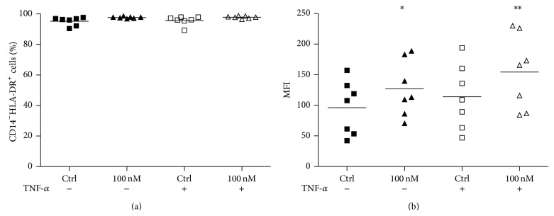
Figure 4.

HLA-DR expression on dendritic cells during activation. After the differentiation period (5 days), cells were incubated for further 48 h with or without TNF-α (50 ng/mL), in the presence or absence of 100 nM Oua. HLA-DR expression was analyzed by flow cytometry. Data are expressed as (a) the percentage of HLA+ cells, or (b) MFI (mean of fluorescence intensity), and lines denote the means of seven independent experiments. ∗ and ∗∗ are significantly different from the control (∗ P < 0.05; ∗∗ P < 0.01).
