
Figure 5.
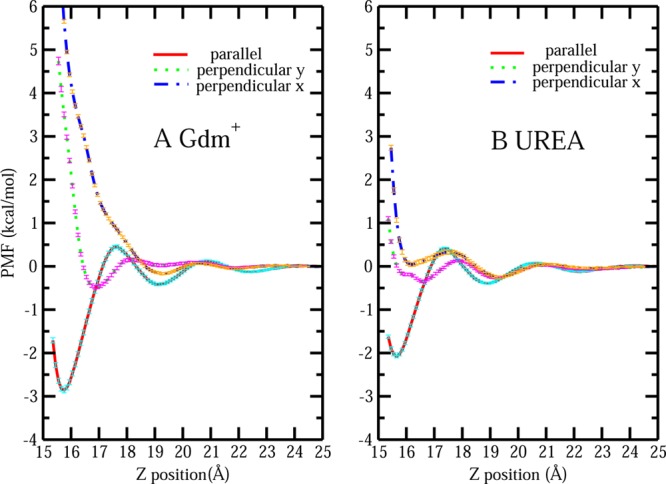
(A) PMF for single Gdm+ with parallel orientation, perpendicular y and perpendicular x orientation from bulk approaching the hydrophobic protein–solvent interface. (B) PMF for single urea with parallel orientation, perpendicular y, and perpendicular x orientation from bulk approaching the hydrophobic protein–solvent interface.