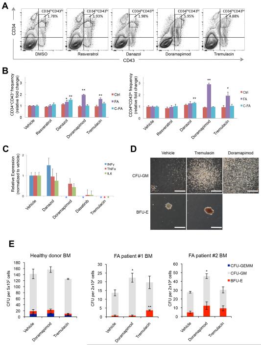
Fig 8. Small-molecule screen for compounds rescuing FA hematopoietic defects.
Two randomly selected clones, FA-iPSC#5 and FA-iPSC#8 (data not shown) were used in this experiment and provided consistent results. A, FACS analysis of the CD34+ and CD43+ populations at day 13 of hematopoietic differentiation of FA-iPSC#5 after one-week treatment with vehicle (DMSO), resveratrol (1 μM), danazol (50 ng/ml), doramapimod (5 μM) and tremulacin (5 nM). B, Quantification of percentages of FA-HPCs that are CD34+/CD43+ and CD34hi/CD43lo after drug treatments indicated in A. Error bars represent SEM of 3 independent experiments. * p<0.05 and ** p<0.01 (t-test). C, RT-qPCR quantification of expression levels of interferon gamma (INFγ), tumor necrosis factor alpha (TNFα) and Interleukin 6 (IL6) in differentiation cultures of FA-iPSCs treated with vehicle (DMSO), danazol (50 ng/ml), doramapimod (5 μM), dasatinib (5 μM) and tremulacin (5 nM). Expression levels are normalized against GAPDH. Asterisks denote expression levels below the detection limit. D-E, Colony forming assay of FA patient BM mononuclear cells treated with compounds. Representative photos of the morphology of different hematopoietic colonies are shown (D). Bar, 500 μm. E, quantification of the indicated colony types derived from a total of 5×10² BM CD34+ cells and 2×10⁴ BM cells from healthy donors and FA patients, respectively. CFU-GEMM, colony-forming unit granulocyte/erythroid/macrophage/megakaryocyte; CFU-GM, colony-forming unit granulocyte/monocyte; BFU-E, blast-forming unit erythroid. Data are shown as mean±s.d. n=3. * p<0.05 and ** p<0.01 (t-test).

