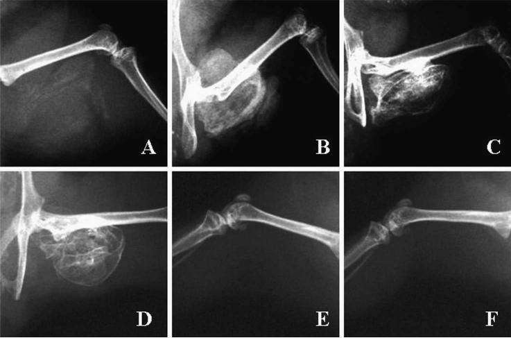
Fig 3.
Radiographic examinations of orthotopic bone in SCID mice hindlimbs are shown. Orthotopic bone formation was seen after implantation of rBMCs transduced with AdBMP-2Myc at (A) At Day 7, orthotopic bone is beginning to form. (B) At Day 14, bone formation has peaked. At (C) Day 21, and (D) Day 56, additional maturation of the orthotopic bone has occurred. Radiographs taken on Day 56 of mice receiving implants containing (E) rBMCs transduced with Adβ-gal or (F) rBMCs alone show no mineralization or orthotopic bone formation.

