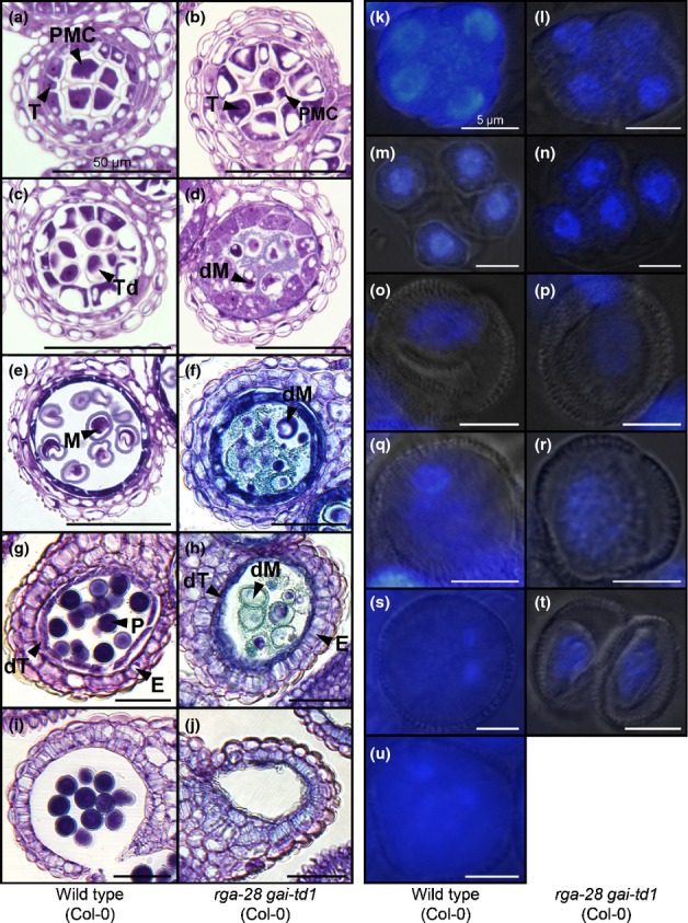
Figure 4.

Microscopic analysis of rga-28 gai-td1 (Arabidopsis thaliana Col-0) pollen development. (a–j) Sections through wild-type (a, c, e, g, i) and rga-28 gai-td1 (Col-0) (b, d, f, h, j) anthers, encompassing developmental stages 5–6 (a, b), 7 (c, d), 8–9 (e, f), 11 (g, h) and 13 (i, j), as defined by Sanders et al. (1999). E, endothecium; dM, degenerating microspore; dT, degenerating tapetum; M, microspore; P, pollen; PMC, pollen mother cell; T, tapetum; Td, tetrad. (k–u) 4′,6-Diamidino-2-phenylindole (DAPI) fluorescence imaging of pollen nuclei in wild-type (k, m, o, q, s, u) and rga-28 gai-td1 (Col-0) (l, n, p, r, t) during development, showing tetrad formation (k–n), free unicellular microspores (o, p), polarized microspores (q), bicellular pollen (s) and mature tricellular pollen (u). rga-28 gai-td1 (Col-0) microspores do not polarize (p), and subsequently degenerate (r, t).
