
Figure 3. IL-22BP staining on sorted subsets of rat splenic conventional DC.
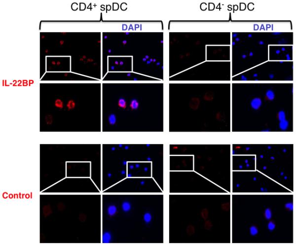
The two subsets of rat conventional spleen DCs were isolated by cell sorting and let to adhere on poly-L-Lysine pre-coated slides for 30 min in the presence of Brefeldin A and monensin. Slides were then stained with a goat anti-rat IL-22BP pAb followed by a donkey anti-goat IgG-Alexa fluor 568. Controls were performed using the secondary antibody alone. Data are representative of two independent experiments.