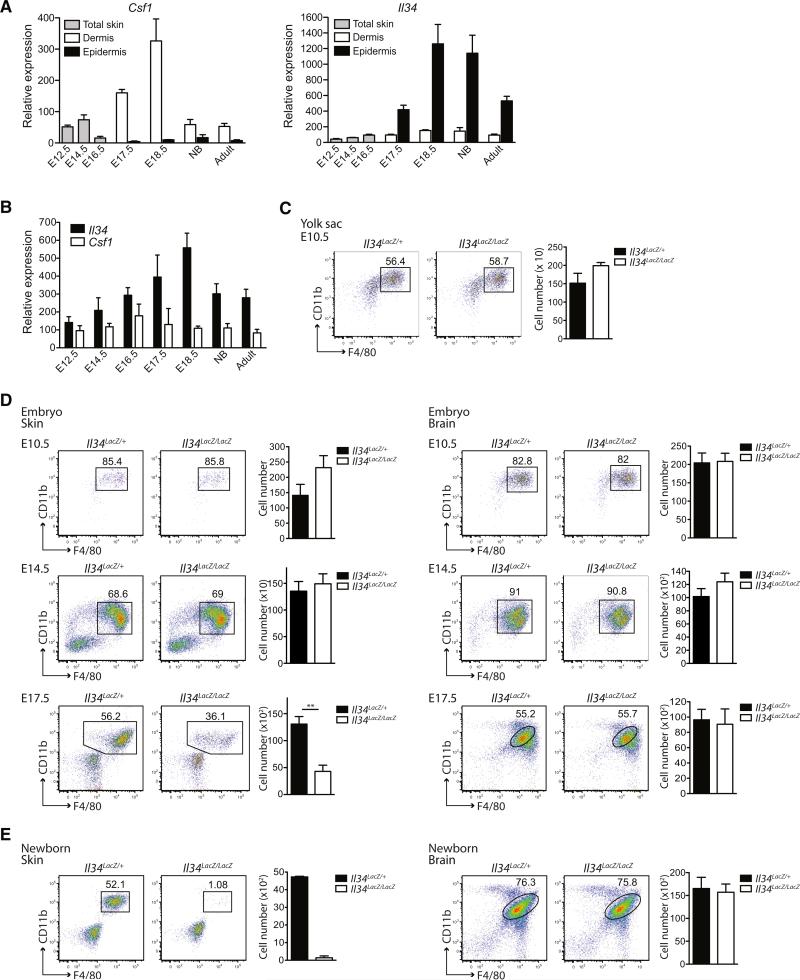
Figure 4. IL-34 Controls the Development of LCs during Embryogenesis.
(A and B) Quantitative RT-PCR analysis of Il34 and Csf1 mRNA expression in the developing skin (A) and the developing brain (B) at different time points during embryogenesis and in newborn (NB) and adult WT mice. Total skin is shown from E12.5–E16.5, and epidermis and dermis are shown after E17.5. Data were normalized to the expression of HPRT1 (n = 3). Shown are pooled data of two individual experiments.
(C) Plots show the percentage (among CD45+ cells) and total cell number (±SEM) of primitive macrophages (CD11b+F4/80+) in the yolk sac at E10.5 (n ≥ 3).
(D) Plots show the percentage and total cell number (±SEM) (among CD45+ cells) of microglia precursors (CD11b+F4/80+) in the brain at E10.5, E14.5, and E17.5 and of LC precursors (CD11b+F4/80+) in the skin (limb buds) at E10.5 and E14.5 and in the epidermis at E17.5 in Il34LacZ/LacZ and Il34LacZ/+ embryos (for E10.5: n ≥ 3, for E14.5: n ≥ 4 and for E17.5: n = 5). Data are pooled of 1–2 different experiments.
(E) Plots show the percentage and total cell number (±SEM) of epidermal LC precursors and microglia (both CD11b+F4/80+) among CD45+ cells in newborn Il34LacZ/LacZ and Il34LacZ/+ mice (LC precursors: n = 2, microglia: n = 6). One representative of three individual experiments is shown. (C–E) *p < 0.05, **p < 0.01, ***p < 0.001 (Student's t test, unpaired).

