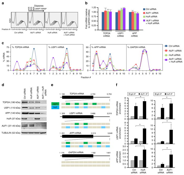
Figure 5. AUF1 cooperates with HuR for mRNA translation.
(a–d) Forty-eight hours after HeLa cells were transfected with the siRNAs indicated, lysates were fractionated through sucrose gradients (a); arrow indicates the direction of sedimentation; −, fractions without ribosomal components, 40S and 60S, small and large ribosome subunits, respectively; 80S, monosome; LMWP and HMWP, low- and high-molecular weight polysomes, respectively. The relative total levels of TOP2A, APP, USP1 mRNAs (normalized to GAPDH mRNA) were assessed (b) and the distribution (%) of TOP2A, APP, USP1 mRNAs and control GAPDH mRNA was measured by RT-qPCR analysis of RNA in each of 10 gradient fractions (c) and the levels of the encoded proteins in whole-cell lysates were assessed by western blot analysis (d). (e) Schematic of AUF1- and HuR-binding sites on the 3′UTRs of TOP2A, APP, USP1 mRNAs and control GAPDH mRNA. (f) Forty-eight hours after silencing HuR or AUF1, the levels of TOP2A, APP, USP1 mRNAs in AUF1 IP or HuR IP were measured by RIP followed by RT-qPCR analysis; data were normalized to control GAPDH mRNA levels in each IP. Data in b,f are the means and s.d. from three independent experiments; *P<0.05, Student’s t-test.

