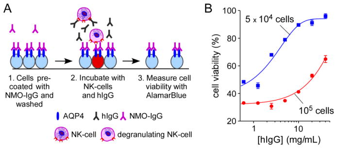
Figure 5. hIgG strongly inhibits NMO-IgG-mediated ADCC.

A. Experimental protocol to measure cytotoxicity produced by NK-cell addition to NMO-IgG-coated CHO-AQP4 cells. CHO-AQP4 cells were incubated with 20 μg/mL rAb-53 for 1 h at 23 °C and washed extensively. NK-cells were pre-incubated with hIgG for 1 h at 37 °C and then added to target cells for 2 h at 37 °C. Target cells were then washed extensively in PBS and viability was measured by addition of 20% AlamarBlue for 1 h at 37 °C. B. Cell viability as a function of hIgG concentration for different numbers of NK-cells added per well (mean ± S.E., n=3).
