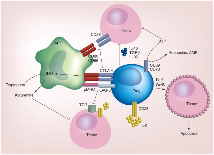
Figure 1. The major cell-contact-dependent and -independent mechanisms utilized by Tregs to suppress conventional T-cell responses.
Tregs express surface receptors, including LAG-3 and CTLA-4, which mediate the cell-contact-dependent suppression of Tconv. These molecules bind pMHC and CD80/CD86, respectively. Subsequently, TCR-pMHC and CD28-CD80/CD86 interactions are disrupted, leading to impaired T-cell activation. CTLA-4-CD80/CD86 interactions also induce APCs to express IDO, which catabolizes tryptophan and therefore reduces the availability of this amino acid needed for T-cell activation. Tregs also produce or respond to soluble factors to suppress Tconv activation. For instance, given their high expression of CD25 relative to Tconv, IL-2 signaling is more robust in Tregs. As a result, there is less IL-2 available to Tconv to promote their activation. Tregs secrete anti-inflammatory cytokines, including IL-10, TGF-β and IL-35 to limit Tconv activation. Tregs that express CD39 and CD73 can deplete a microenvironment of ATP by generating adenosine and AMP, which have immunosuppressive effects on Tconv. Under certain conditions, Tregs may also elaborate Perf and GrzB to induce apoptosis of Tconv. Other surface receptors, including Nrp1, CD103 and ICOS, play vital roles in mediating Treg suppression, but are not depicted here.
GrzB: Granzyme B; Perf: Perforin; pMHC: Peptide-MHC; Tconv: Conventional T cell; TCR: T-cell antigen receptor.

