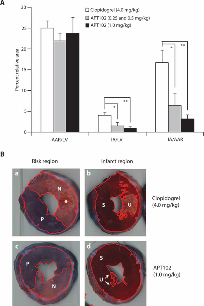
Fig. 5. Effect of clopidogrel and APT102 on myocardial ischemic damage in dogs.
(A) Area at risk (AAR) and infarcted area (IA; nonviable) after coronary artery occlusion of left ventricular (LV) heart muscle of dogs (n = 6) expressed as a percent of the total LV and as a ratio (29). (B) Photographs of representative LV slices from two hearts at the same level showing (left) similar AAR after clopidogrel (a) or high-dose APT102 (c) defined by the area that was nonperfused (N) by Evans blue dye compared to the total surface area including the area perfused (P) with dye. IA, defined as unstained (U) tissue after incubation with triphenyltetrazolium chloride (TTC), was compared to the total area including still viable TTC-stained (S) myocardium for the dog given APT102 (d, arrows) compared to the area in the dog given clopidogrel (b). Infarction in clopidogrel-treated hearts was sometimes marked by more abundant areas of hemorrhage seen in the pre-TTC–stained tissue (*, panel a). Bars represent means ± SEM. *P < 0.02, **P < 0.002 by t test.

