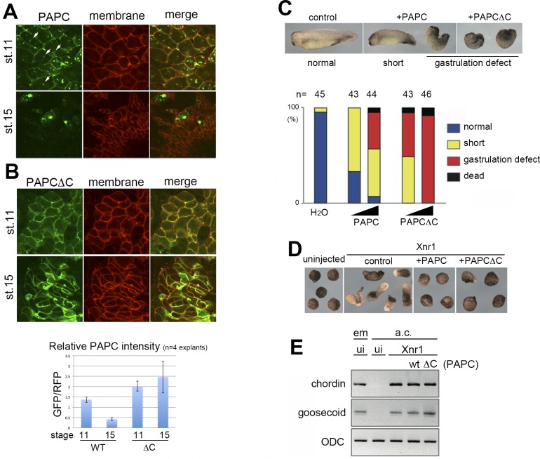
Figure 1. PAPC is down-regulated in the dorsal marginal zone explants at late gastrula.
(A) GFP-tagged PAPC (PAPC-GFP) was expressed in the dorsal marginal zone (DMZ) of a Xenopus embryo. In the DMZ explant at stage 11 (early gastrula), PAPC-GFP was found mainly on the plasma membrane (marked by membrane-targeted RFP), with some cytoplasmic puncta (arrowheads). At stage 15 (early neurula), PAPC-GFP signal was greatly reduced and excluded from the plasma membrane. (B) GFP-tagged PAPC mutant lacking the cytoplasmic domain (PAPCΔC) was not degraded and persistently localized to the plasma membrane at stages 11 and 15. PAPC-GFP signal was reduced at stage 15 as measured by relative fluorescent intensity using membrane-RFP as an internal control, while PAPCΔC-GFP signal persisted. (C) Injecting an increasing amount of wild-type PAPC mRNA into Xenopus embryos induced more severe gastrulation defects, including a short body axis and spina bifida at stage 28. PAPCΔC over-expression exhibited stronger effects. The amount of injected RNAs was 200 and 1000 pg, respectively. (D) Over-expression of PAPC or PAPCΔC inhibited elongation of animal caps mesodermalized by Xnr1 expression. (E) Induction of mesoderm was confirmed by expression of chordin and goosecoid (gsc) by RT-PCR. em, embryos; a.c., animal caps; ui, uninjected.

